

l

Crumpled cytoplasm, clear diagnosis: the iconic morphology of Gaucher disease
#00066595
Author: Derek C. Sung; Dale M. Frank
Category: Myeloid Neoplasms and acute leukemia (WHO 2016) > Myelodysplastic/myeloproliferative neoplasms (MDS/MPN)
Published Date: 08/20/2026
Category: Myeloid Neoplasms and acute leukemia (WHO 2016) > Myelodysplastic/myeloproliferative neoplasms (MDS/MPN)
Published Date: 08/20/2026
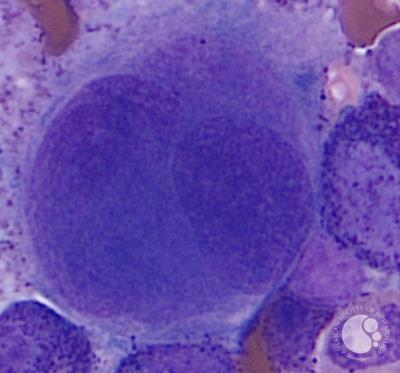
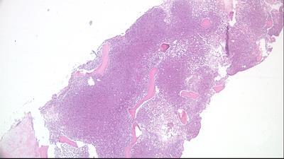
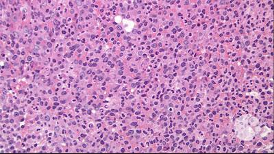
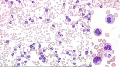
A 22-year-old woman initially presented with infectious mononucleosis and thrombocytopenia. Serial laboratory studies over 2 years showed persistent thrombocytopenia with platelets ranging from 74 × 109/L to 104 × 109/L. Ultrasound imaging and magnetic resonance imaging revealed hepatosplenomegaly and splenic lesions. The patient underwent bone marrow studies with an aspirate revealing abundant histiocytes with fibrillary “crumpled tissue paper” cytoplasm (panel A; Wright-Giemsa stain; 60× objective), consistent with Gaucher cells, and a biopsy showing sheets of Gaucher cells (panel B; hematoxylin and eosin stain; 20× objective) positive for CD68 by immunohistochemistry (panel C; 20× objective). Additional testing on peripheral blood revealed elevated glucosylsphingosine and decreased leukocyte β-glucosidase activity. Targeted long-range polymerase chain reaction and next-generation sequencing on
The p.P358L mutation has been reported only twice in GD, both compound heterozygotes. Type 1 GD can present in childhood or adulthood with anemia, thrombocytopenia, hepatosplenomegaly, and bone/joint pain, and, in contrast to type 2 and 3 disease, it lacks neurologic symptoms. Although marrow involvement is common, diagnosis requires only genetic or enzymatic studies performed on peripheral blood. GD is prevalent among the Ashkenazi Jewish population, with a carrier rate of 1 in 14. The patient has no known Ashkenazi Jewish ancestry, although her maternal ancestry is uncertain because the patient’s mother was adopted.
For additional images, visit the ASH Image Bank, a reference and teaching tool that is continually updated with new atlas and case study images. For more information, visit


Related Images: