

l

Disseminated cryptococcal lymphadenitis mimicking lymphoma and masked by prozone effect
#00066623
Author: Andrew W. Allbee; Sam Sadigh
Category: Myeloid Neoplasms and acute leukemia (WHO 2016) > Myelodysplastic/myeloproliferative neoplasms (MDS/MPN)
Published Date: 08/20/2026
Category: Myeloid Neoplasms and acute leukemia (WHO 2016) > Myelodysplastic/myeloproliferative neoplasms (MDS/MPN)
Published Date: 08/20/2026
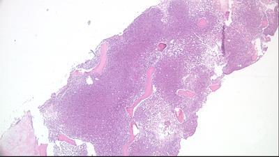
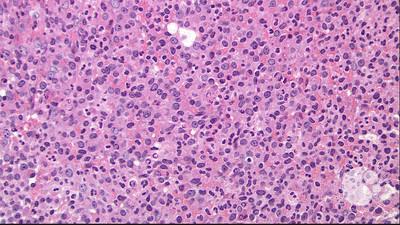
A 55-year-old man with no medical history presented with weakness, weight loss, and drenching night sweats. A complete blood count showed mild anemia (hemoglobin, 11.3 g/dL) but was otherwise normal. Computed tomography demonstrated multistation thoracic/abdominal lymphadenopathy (up to 3.0 cm), raising suspicion for lymphoma. However, retroperitoneal lymph node biopsy revealed replacement by histiocytes and innumerable yeast forms with clear halos, refractile capsules, and narrow-based budding (panels A-C; hematoxylin and eosin; 4×, 50×, and 100× objectives, respectively). Periodic acid–Schiff, Grocott-Gomori methenamine silver, and mucicarmine stains were positive (panels D-F; 100× objective). Serum cryptococcal antigen was initially negative due to the prozone effect, also known as the hook effect, a condition in which antigen overabundance oversaturates reagents and leads to a false-negative result. However, high suspicion prompted a dilution study that confirmed a markedly positive titer >1:2560. Cerebrospinal fluid studies also showed India ink–positive cryptococci. Further workup revealed
Although the incidence of AIDS as the initial presentation of HIV infection is now rare in areas with access to screening/ART, this exceptional case of disseminated cryptococcosis serves as an instructional reminder to include opportunistic infections in the differential diagnosis for lymphadenopathy.
For additional images, visit the ASH Image Bank, a reference and teaching tool that is continually updated with new atlas and case study images. For more information, visit


Related Images: