

l

Blasting into muscle: isolated BPDCN relapse with second TP53 hit
#00066648
Author: Federico Repetto; Sam Sadigh
Category: Myeloid Neoplasms and acute leukemia (WHO 2016) > Myelodysplastic/myeloproliferative neoplasms (MDS/MPN)
Published Date: 08/20/2026
Category: Myeloid Neoplasms and acute leukemia (WHO 2016) > Myelodysplastic/myeloproliferative neoplasms (MDS/MPN)
Published Date: 08/20/2026
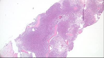
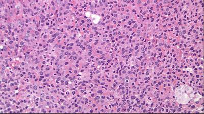
An 81-year-old man with blastic plasmacytoid dendritic cell neoplasm (BPDCN) involving the skin, bone marrow, and central nervous system achieved complete remission with pivekimab sunirine, intrathecal chemotherapy, and allogeneic stem cell transplant. After a localized cutaneous relapse treated with radiation and donor lymphocyte infusion, surveillance positron emission tomography/computed tomography obtained during follow-up for metastatic prostate cancer showed a new intensely fluorodeoxyglucose-avid, weakly prostate-specific membrane antigen-avid intramuscular left thigh lesion (panel A) without active skin disease. Biopsy showed skeletal muscle diffusely replaced by medium/large-sized blasts with irregular nuclei and fine chromatin (panels B-C; hematoxylin and eosin; original magnification ×4 and ×100, respectively). By immunostaining, the cells expressed TCF4/CD123, TCL1, and SOX4 (panels D-F; original magnification ×100) and showed diffuse p53 overexpression (panel G; original magnification ×100). At initial diagnosis, next-generation sequencing had revealed canonical founder alterations with biallelic
This case highlights an unusual intramuscular relapse of BPDCN without active skin disease and illustrates stepwise
For additional images, visit the ASH Image Bank, a reference and teaching tool that is continually updated with new atlas and case study images. For more information, visit


Related Images: