

l

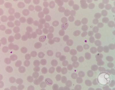
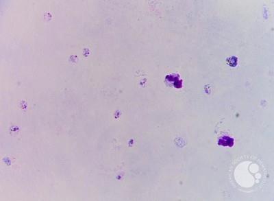
Trophozoites of P.vivax
#00066739
Author: Gurpreet Kaur, MD,DM; Ankur Ahuja, MD.,DM; Anu R.; PadmaPrakash ,MD.DM
Category: Infectious Disease > Parasites > Malaria > P. vivax
Published Date: 10/15/2026
Category: Infectious Disease > Parasites > Malaria > P. vivax
Published Date: 10/15/2026
P. vivax developing trophozoites exhibit an irregular, amoeboid cytoplasm that looks active, spreading across the cell with prominent pseudopodial extensions. As the trophozoite matures, fine, yellowish-brown to golden-brown malarial pigment (hemozoin) accumulates and spreads within the cytoplasm.P. vivax preferentially infects reticulocytes; consequently, the infected RBC is significantly larger and paler than the surrounding uninfected cells. The cytoplasm of the infected erythrocyte displays fine, evenly distributed pink/red stippling known as Schüffner’s dots.


Related Images: