

l

Optical genome mapping reveals a MYC-amplified ring chromosome arising through chromoanagenesis in AML
#00066754
Author: Guilin Tang; Ying S. Zou
Category: Myeloid Neoplasms and acute leukemia (WHO 2016) > Myelodysplastic/myeloproliferative neoplasms (MDS/MPN)
Published Date: 10/15/2026
Category: Myeloid Neoplasms and acute leukemia (WHO 2016) > Myelodysplastic/myeloproliferative neoplasms (MDS/MPN)
Published Date: 10/15/2026
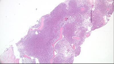
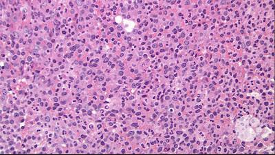
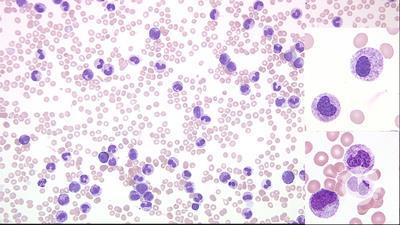
An 82-year-old man presented with progressive dyspnea. Peripheral blood showed leukocytosis (white blood cell, 80 × 109/L), anemia (hemoglobin, 8.9 g/dL), thrombocytopenia (platelets, 31 × 109/L), and 35% blasts (panel A, Wright-Giemsa stain, 100× objective). Bone marrow was hypercellular (90%) with 70% blasts (panel B, hematoxylin and eosin stain, 40× objective). The blasts were large with moderate cytoplasm, and some had cytoplasmic vacuoles and/or Auer rods (panel C, Wright-Giemsa stain, 100× objective). Flow cytometry identified a CD45-dim blast population (32%) expressing CD33, CD117, CD123, HLA-DR, and myeloperoxidase, with partial expression of CD4, CD13, CD34, and CD64. The patient was diagnosed with acute myeloid leukemia (AML). Conventional cytogenetics revealed 47,XY,+r[20]. Fluorescence in situ hybridization using a
Ring chromosomes can serve as large carriers of amplified extrachromosomal DNA harboring oncogenes. OGM resolved the complex genomic architecture of a
For additional images, visit the ASH Image Bank, a reference and teaching tool that is continually updated with new atlas and case study images. For more information, visit


Related Images: