

l

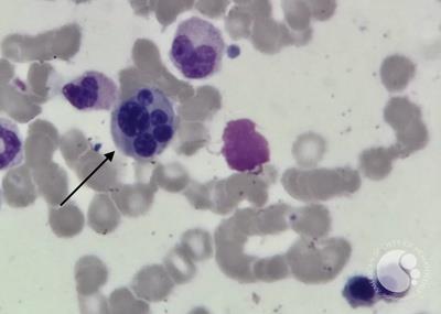


Figure 2: Leishman–Giemsa-stained bone marrow aspirate smear demonstrates prominent dyserythropoiesis
#00066603
Author: Shilpi Saxena, MD; VA Arun
Category: Red Cell: Disorders of Iron Metabolism and Heme Synthesis > Sideroblastic anemias > Acquired/Reversible Sideroblastic Anemias > Myelodysplastic Syndromes
Published Date: 08/21/2026
Category: Red Cell: Disorders of Iron Metabolism and Heme Synthesis > Sideroblastic anemias > Acquired/Reversible Sideroblastic Anemias > Myelodysplastic Syndromes
Published Date: 08/21/2026
An elderly male presenting with refractory anaemia and transfusion-dependent for the past 6 months. Complete blood count revealed pancytopenia. Peripheral blood smear revealed macrocytic anaemia with grade 3 neutropenia and dysmyelopoiesis (pseudo Pelger-Huet neutrophils; Figure 1). The bone marrow aspirate smears was hypercellular with trilineage hematopoiesis with dyserythropoiesis in the form of binucleation / multinucleation, nuclear budding, internuclear bridging, irregular nuclear contours, karyorrhexis and megaloblastoid changes in >10% erythroid precursors (Figures 2,3). Dysmegakaryopoiesis was also present (Figure 4). Blasts constituted <5% of nucleated cells. The provisional diagnosis of myelodysplastic neoplasm with multilineage dysplasia was offered. These findings must be interpreted with clinical context and cytogenetics to exclude mimics (e.g., megaloblastic anemia, drug effect)
Myelodysplastic syndromes (MDS) are clonal hematopoietic neoplasms characterized by ineffective hematopoiesis, cytopenias, and morphologic dysplasia, with a risk of progression to acute myeloid leukemia.
Morphologic dysplasia remains central to diagnosis, requiring ≥10% dysplastic cells in a lineage for significance.


Related Images: