

l

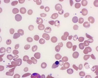
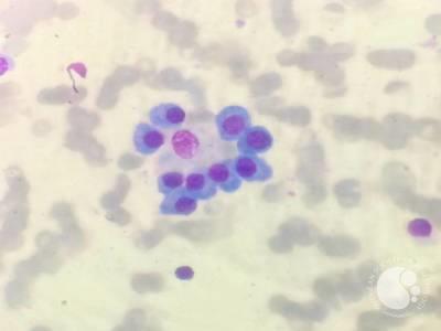
Figure 2: Bone marrow aspirate smears showing numerous hemosiderin-laden macrophages with coarse blue granules, consistent with Grade 6 iron stores ( (Perls’ Prussian blue stain)
#00066607
Author: Shilpi Saxena, MD; Priyanka Mishra; VA Arun
Category: Red Cell: Disorders of Iron Metabolism and Heme Synthesis > Hemochromatosis > x linked (and other) hereditary sideroblastic anmia > Multiple blood transfusions
Published Date: 08/21/2026
Category: Red Cell: Disorders of Iron Metabolism and Heme Synthesis > Hemochromatosis > x linked (and other) hereditary sideroblastic anmia > Multiple blood transfusions
Published Date: 08/21/2026
A transfusion-dependent patient with Thalassemia presents with chronic anemia and secondary iron overload due to repeated red cell transfusions.
Leishman Giemsa-stained smears revealed hemosiderin-laden macrophages (Figure 1) containing abundant coarse, refractile, golden-brown cytoplasmic granules, representing iron in the form of hemosiderin. The granules are variably sized, often clumped, and may partially obscure the macrophage nucleus. Although LG stain does not specifically highlight iron, the dense brown granular pigment within macrophages in the appropriate clinical setting is highly suggestive of marked iron overload.
Bone marrow aspirate smears stained with Perl’s Prussian blue demonstrate the marrow fragments densely packed with iron, which is noted in virtually all fragments examined. Extracellular iron deposition is also seen, indicating markedly increased iron stores (Grade 6/6; Figure 2). Overall findings were consistent with severe iron overload due to chronic transfusion therapy.


Related Images: