

l

Erythrophagocytosis by neoplastic cells in HSTCL
#00066618
Author: Gurpreet Kaur, MD,DM; Ankur Ahuja , MD, DM; Shipra Verma,MD; Ashok Meshraam, MD, DM
Category: Lymphoma: Mature T and NK cell lymphoproliferations > Mature T-cell Lymphomas > Gastro-intestinal T-cell lymphomas > Hepatosplenic T-cell Lymphoma
Published Date: 08/21/2026
Category: Lymphoma: Mature T and NK cell lymphoproliferations > Mature T-cell Lymphomas > Gastro-intestinal T-cell lymphomas > Hepatosplenic T-cell Lymphoma
Published Date: 08/21/2026
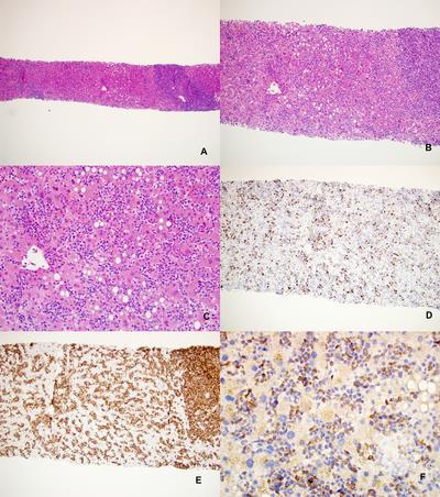
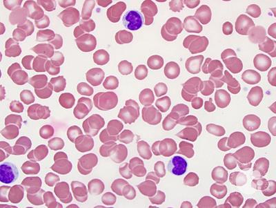
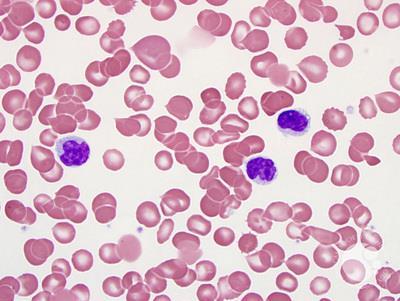
Erythrophagocytosis by neoplastic cells is an extremely rare and distinct cytological finding in Hepatosplenic T-cell lymphoma (HSTCL), as it is more commonly associated with monocytic acute myeloid leukemia (AML). While HSTCL typically presents as mature lymphoid cells, in advanced stages, the cells can exhibit a larger, "blastic" morphology and, on occasion, directly participate in phagocytosis.The malignant cells are generally small to intermediate in size but can transform into larger, blastic-appearing cells that resemble lymphoblasts. We present the case of a 23 years old male with one month history of fever of and loss of appetite. He had pallor with mild hepatomegaly and marked splenomegaly.No lymphadenopathy was seen. Initial laboratory investigations revealed pancytopenia with a hemoglobin level of 7.6 g/dL, a total leukocyte count of 3,800 /µL and platelets of 55,000/µL. Peripheral smear demonstrated microcytic hypochromic red cells, leucopenia with left shift and thrombocytopenia with blastoid cells with erythrophagocytosis.Figure 1.A,B Bone marrow (BM) aspirate smears revealed cellular marrow with atypical blastoid cells (42%) which were small to medium size with pale basophilic cytoplasm, and minimally condensed chromatin with one to two prominent nucleoli. The bone marrow biopsy revealed sheets of atypical cells along with reduced trilineage hematopoietic elements. Flow cytometric analysis of the BM aspirate revealed the presence of approximately 38% abnormal T cells within the presumptive lymphocyte gate (bright CD45 with low side scatter) expressing bright sCD3 dim CD5 and negative CD56 expression. These cells were negative for cytoplasmic CD4, and CD8 and expressed TCR gamma delta. The cells did not express immaturity markers (CD34, CD117, TdT) or B-lymphoid or myeloid markers. Based on this a diagnosis of Hepatosplenic T-Cell Lymphoma was rendered and the patient was started on therapy and planned for Hematopoietic stem cell transplantation.
It is crucial to distinguish this from AML on morphology specifically monoblastic leukemia with t(8;16)), which often shows blastic erythrophagocytosis. In contrast, HSTCL is typically CD3+/CD56+ and CD4-/CD8- (double negative), whereas AML is CD34/CD117 positive.While the exact mechanism is unknown, it is hypothesized that the tumor cells become activated and exhibit phagocytic capabilities, perhaps due to cytokine production.The presence of blastic erythrophagocytosis is typically a sign of advanced, highly aggressive disease with a very poor prognosis, often indicating resistance to conventional chemotherapy.Although standard treatments often fail, HSCT has been reported as a potentially curative option.
Figure1.Peripheral blood smear with Erythrophagocytosis by blastoid cells with dispersed nuclear chromatin, multiple inconspicuous nucleoli, and occasional vacuolated cytoplasm (Leishman Giemsa stain 100X)


Related Images: